

- Cet évènement est passé.
Des images de microscopie aux modèles physiques : décrypter la morphogenèse des tissus
Hervé Turlier (CBI)
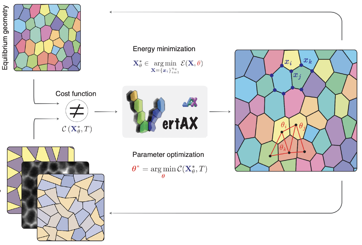
Fluorescence microscopy now gives detailed 2D and 3D images of tissues and embryos, yet turning these images into physical quantities such as forces, tensions or material constants is still hard. In this talk I will approach the question as an inverse problem. I will present VertAX, a differentiable 2D vertex model in which the tissue geometry is a mechanical equilibrium, and in which cell–cell tensions are learned by bilevel optimization. The images turn out to contain the information we need; the difficulty comes from the equilibrium itself, whose soft modes convert small errors in vertex positions into large errors in forces. I will show how simple physical arguments help, for instance choosing the right metric or stopping the relaxation early, and where the Cramér–Rao bound sets a limit that no unbiased estimator can beat. The same framework can also be used to design tissue shapes and mechanical properties. I will end with perspectives on 3D tissues, dissipative dynamics, and reinforcement learning for situations, such as cell divisions, where the problem is no longer differentiable.
Contact : N. Destainville